
Symptom library

Spots on Skin — Identify by Body Area

By ScanSkinAI Editorial Team Reviewed for medical safetyLast reviewed:

What is this spot on my skin? Pick the body area below to see the most likely causes for red, brown, white, black, pearly or itchy spots on that exact location, the red-flag warning signs of skin cancer, and clear advice on whether to monitor at home or book a dermatology review. 480 plain-English guides, reviewed against current dermatology guidance.
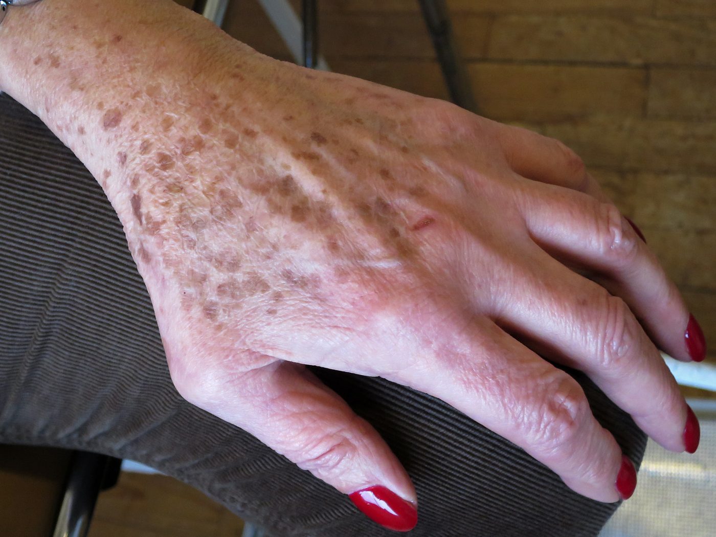
Clinical photo of a solar lentigo (age spot) — a flat, uniformly pigmented brown spot on sun-exposed skin.
Solar lentigo (age spot) — a flat, uniformly pigmented brown patch of sun-damaged skin, one of the most common adult skin spots. Image: Wikimedia Commons · Alain Gérard · CC BY-SA 4.0

Identify a skin spot by colour

Not sure where to start? Jump to the colour that matches the spot you're seeing — each link opens a dedicated guide with photos, likely causes and red-flag warning signs.

Identify a skin spot by body area

Different parts of the body see different sun exposure, friction and sweat patterns — so the same-looking spot can mean different things on the face versus the foot. Pick the location closest to your spot.

Prefer to search by condition name, symptom or rash type instead of body area? These hubs cover the same library from different angles.

Spots on skin — frequently asked questions

Most adult skin spots fall into a handful of common groups: sun spots (solar lentigines) and seborrhoeic keratoses (benign brown growths), cherry angiomas (small red dots), milia and whiteheads (white bumps), folliculitis and acne (red bumps), and skin tags. The minority that matter most are basal cell carcinoma (pearly bump that won't heal), squamous cell carcinoma (rough scaly patch), and melanoma (asymmetric, multi-coloured or changing mole). Pick the body area above to see the most likely causes for spots in that exact location.

Use the ABCDE rule for pigmented spots: Asymmetry, irregular Borders, more than one Colour, Diameter over 6 mm, and Evolution (any change in size, shape or colour). For non-pigmented skin cancer, watch for a pearly bump with visible tiny blood vessels (basal cell), a scaly or crusty patch that won't heal in 4 weeks (squamous cell or actinic keratosis), or any sore that bleeds repeatedly. When in doubt, a free AI mole check gives an instant risk indication.

Brown spots are usually pigment-related — sun spots, freckles, seborrhoeic keratoses, melasma or moles. Red spots are most often inflammation (acne, folliculitis, eczema), broken blood vessels (cherry angiomas, petechiae) or allergic rashes. White spots are typically pigment loss (vitiligo, pityriasis alba), fungal infection (tinea versicolor) or sun-damage-related (idiopathic guttate hypomelanosis). Each colour has its own dedicated guide linked above.

Book a dermatology review within 2–4 weeks if a spot is new in adulthood, changing in size, shape or colour, bleeding or crusting without obvious trauma, asymmetric or multi-coloured, larger than 6 mm, or the 'ugly duckling' that looks different from your other moles. Seek same-day care for spreading redness with fever, a painful purple rash that doesn't blanch, or any rapidly growing painful lump.

Photograph the spot in even daylight with a coin or ruler next to it for scale, then upload it to the free AI mole checker. The tool gives an instant ABCDE-style risk indication and lets you compare the same spot over weeks or months to detect change — the single strongest signal of an early skin cancer.

We publish 480 plain-English guides covering 12 body locations (face, scalp, neck, chest, back, abdomen, arm, hand, leg, foot, groin, buttock) crossed with 40 common spot types — red, white, brown, black, pearly, scaly, itchy, painful, bleeding, growing and more. Each guide is reviewed against current dermatology guidance.