
Full-Body Mole Mapping at Home: A Smarter Way to Track 50+ Moles
If you have more than 50 moles, memory and phone photos are not enough. A structured mole map turns hundreds of individual moles into a system you can actually monitor — so genuine change becomes obvious, fast.
Most users scan once and feel reassured. The ones who catch problems early scan every 4–6 weeks and compare results over time.
Most users scan once and feel reassured. The ones who catch problems early scan every 4–6 weeks and compare results over time.
Quick answer
Full-body mole mapping is a structured way to photograph and record every mole on your body so that new, growing, or changing moles stand out over time. For people with 50+ moles, it is the single most effective at-home practice for early melanoma awareness — but it is a screening tool, not a diagnosis. Any flagged mole should be reviewed by a dermatologist.
Why people with 50+ moles can't track moles reliably at home
Most people with high mole counts only realise the scale of the problem when a dermatologist says: "Has this one always looked like that?"Without a baseline, the honest answer is usually "I don't know."
The "which one changed?" problem
When you have 80, 100, or 200 moles, you cannot hold them all in memory. A new mole hides among existing ones. A subtly enlarging mole looks identical to what you already had. Change is invisible without a reference image.
Why memory and phone photos fail
Random camera-roll shots have inconsistent lighting, distance and framing. They are not tagged by body location. Six months later you cannot find the photo of that mole on the lower back — and even if you do, you cannot reliably compare it.
What full-body mole mapping is
Mole mapping is the practice of dividing the body into zones, photographing each zone in a repeatable way, and tagging every individual mole to a location on a body diagram. Repeat the scan consistently and any change — new mole, growing mole, colour shift — becomes obvious by comparison.
Body zones & numbering basics
A standard mole map divides the body into roughly 12 zones (face, scalp, front chest, abdomen, upper back, lower back, each arm, each leg, hands, feet). Each mole is tagged to a zone and given a persistent ID so it can be re-photographed over time.
Baseline vs. follow-up scans
Your baseline is the first complete scan — every zone, every mole. Follow-up scans repeat the same zones every 1–3 months. The system compares against the baseline so you do not have to rely on memory.
What "change" actually means
Change is judged using the ABCDE rule: Asymmetry, Border irregularity, Colour variation, Diameter over 6 mm, and — most importantly — Evolving. Evolution over weeks to months is the single most reliable melanoma signal, and it is exactly what a mole map surfaces.
How ScanSkinAI builds your personal mole map
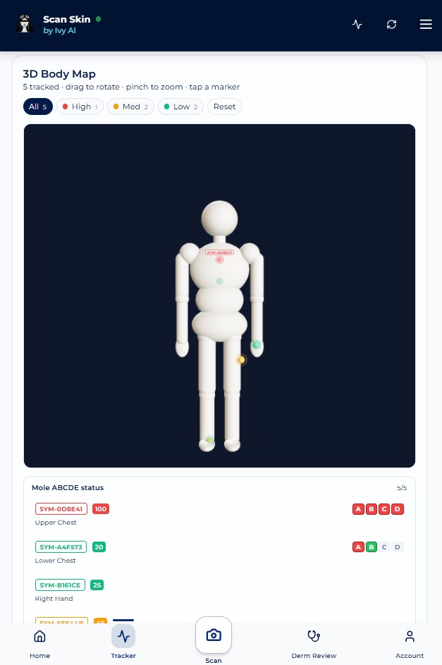
3D body mole map by location
Tag every mole to a 3D body diagram so each one has a fixed address you can return to.
Single-mole timeline tracker
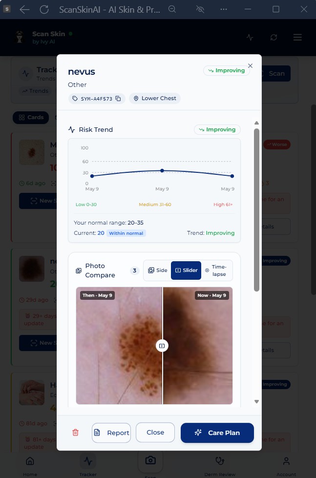
Every individual mole keeps its own dated photo timeline so you can scroll back through every previous scan.
Side-by-side change comparison
Compare any two scans of the same mole frame-by-frame. Subtle growth or colour shift becomes immediately visible.
Never miss a mole check
Set personalised reminders for your next full-body scan and track every mole on schedule.
Who should map their moles
- Anyone with more than 50 moles on the body
- Family history of melanoma or atypical moles
- Fair skin (Fitzpatrick I–II), red or blond hair, light eyes
- History of severe or blistering sunburn, especially in childhood
- Prior personal history of any skin cancer or biopsied atypical mole
- Long-term immunosuppression (e.g. organ transplant, biologics)
- Outdoor workers and frequent UV exposure
Step-by-step: building your baseline at home
- Set the scene. Use bright, even, natural daylight. Plain background. Phone held about 30 cm from the skin for close-ups.
- Work zone by zone. Face → scalp → front torso → back → arms → hands → legs → feet. One wide shot per zone, plus close-ups of any individual mole.
- Tag each mole to its body location. ScanSkinAI's 3D body map does this in one tap.
- Save the baseline. This is your reference set. Everything future is compared against it.
- Set a 3-month recurring reminder to re-scan the same zones in the same way.
When a mole needs professional review
Red-flag signs
- A genuinely new mole appearing after age 40
- Any mole that has grown, darkened, or changed shape between scans
- Two or more colours within a single mole (brown + black, red, blue or white)
- Irregular, notched or fading borders
- A mole that itches, bleeds, scabs, or fails to heal
- A mole that looks visibly different from your other moles ("ugly duckling")
How to bring your map to a doctor
Export the dated photo timeline of the flagged mole from your ScanSkinAI account and bring it to your GP or dermatologist. A clear before/after image set is often enough to shortcut the consultation and prioritise a biopsy.
Mole mapping vs chronic skin tracking
Mole mapping is built for discrete pigmented lesions — individual moles, each with its own timeline. If you are tracking a chronic skin condition instead (eczema, psoriasis, rosacea, acne, vitiligo), the right tool is the Chronic Skin Condition Tracker. It records flare-ups, treatments and severity over time, rather than individual lesions on a body map.
Many users use both: the mole map for melanoma awareness, the chronic tracker for ongoing skin conditions.
Why one check is rarely enough
A single scan tells you about one spot, on one day. But skin changes are about patterns over time — a new mole appearing, a slow shift in shape, size or colour, or a patch that simply isn't healing. Monitoring the same spots side-by-side, week after week, surfaces the subtle changes a one-off check will always miss — and gives you a clear record to show a clinician if something needs a closer look.
(ScanSkinAI is a screening and monitoring tool, not a diagnosis. Always see a clinician for anything that is changing, bleeding, or worrying you.)
Unlimited scans · Track changes side-by-side · Cancel anytime
Related reading: monthly mole-tracking routine
Frequently asked questions
Frequently Asked Questions
Full-body mole mapping is the practice of systematically recording every mole on your body — typically by body zone — so that any new or changing mole can be detected over time. Traditionally done by dermatologists with specialised cameras, it can now be performed at home using smartphone photos plus a digital mole map.
Having more than 50 moles is one of the strongest known risk factors for melanoma. With so many moles, memory and casual phone photos are unreliable — you cannot tell which one is new or which one has changed. A structured mole map gives every mole a location and a baseline image, so change becomes visible.
Yes. You will not get the dermatoscopic detail of a clinic, but for change-detection — which is what catches most early melanomas — a consistent, well-lit photo of each body zone, tagged by location, is highly effective. ScanSkinAI builds the map for you and lets you compare any single mole side-by-side over time.
Set a baseline now and re-scan every 3 months. People with very high mole counts, fair skin, family history of melanoma, or prior atypical moles should re-scan monthly during high-UV seasons. Any mole that changes between scans warrants a closer look.
Use the ABCDE rule: Asymmetry, Border irregularity, Colour variation, Diameter over 6 mm, and Evolving (any change in size, shape, colour, height or sensation). Evolution is the single most important signal — and it is exactly what a mole map is designed to surface.
No. ScanSkinAI is an at-home screening and tracking tool — not a diagnosis. If your map flags a mole as new, changed, or visually atypical, take that record to a dermatologist. The map shortens the consult and helps your clinician spot what changed.
Yes. Your mole map, photos and timeline are stored in your private ScanSkinAI account. They are never shared without your consent and are protected under ISO 27001 information-security controls.
Key takeaways
- Having 50+ moles is one of the strongest known melanoma risk factors.
- Memory and casual phone photos are not enough to detect change at this scale.
- Full-body mole mapping divides the body into zones and tags every mole to a location.
- A 3-month re-scan cadence makes new or evolving moles visible by comparison.
- The ABCDE rule — especially Evolving — is the standard for judging change.
- ScanSkinAI provides a 3D body map, per-mole timeline, and side-by-side comparison at home.
- Any flagged mole should be confirmed by a dermatologist — the map is screening, not diagnosis.
Have more than 50 moles?
Start building your personal mole map today. Track each mole by body location, compare changes over time, and know when a mole may need medical review.
Start Your Mole MapNot sure where to start?
Upload one photo of your most-watched mole. ScanSkinAI screens it free in seconds — then anchors it on your body map so you can track it from here on.