
Basal Cell Carcinoma: Pictures, Symptoms & Treatment
Basal cell carcinoma (BCC) is the most common cancer in humans, with more than four million US cases diagnosed each year. It grows slowly, almost never spreads, and is highly curable — but it keeps growing locally until it is treated.
Quick answer
Basal cell carcinoma typically looks like a pearly or waxy bump with fine surface blood vessels, or a sore that scabs, heals and comes back in the same spot. It is most common on the face, ears, neck, scalp and shoulders. It rarely spreads but is locally destructive, so it needs treatment — usually surgical excision, or Mohs surgery for facial and high-risk lesions.
One free scan gives a first answer — the 3-month plan tracks a slow-growing lesion over time.
Basal cell carcinoma pictures
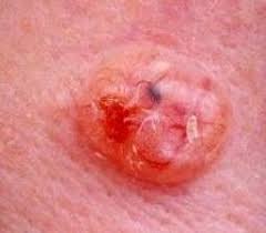
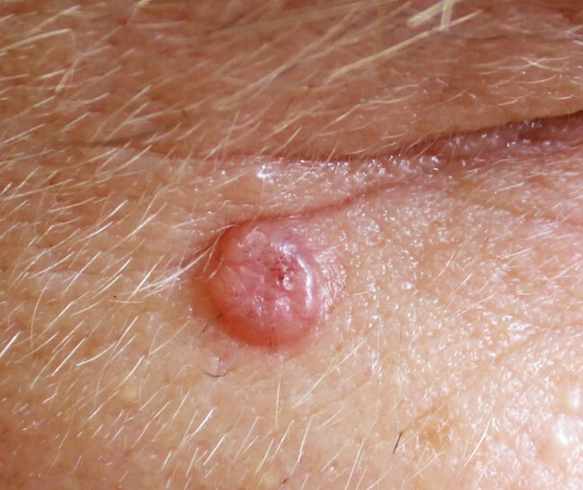
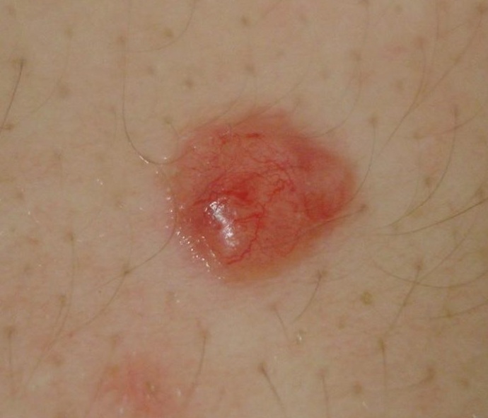
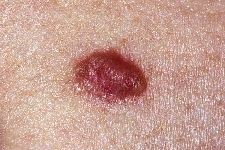
See more examples in our skin cancer pictures guide.
Early signs and symptoms
Common locations
- Nose, cheeks and forehead
- Around the eyes and eyelids
- Ears and behind the ears
- Neck, scalp and shoulders
- Upper back and chest
Warning signs
- Pearly, waxy or translucent bump
- Visible fine blood vessels on the surface
- A sore that bleeds, scabs, then returns
- Flat, scar-like patch with no injury history
- Pink growth with a rolled edge and crusted centre
BCC subtypes
| Subtype | Appearance and behaviour |
|---|---|
| Nodular | Most common. Pearly dome-shaped papule with telangiectasia, often ulcerating in the centre. |
| Superficial | Flat pink or red scaly patch, frequently on the trunk. Easily mistaken for eczema or psoriasis. |
| Morpheaform (sclerosing) | Waxy, scar-like plaque with poorly defined edges. More aggressive; usually needs Mohs surgery. |
| Pigmented | Contains brown or black pigment and can be confused with melanoma. |
Causes and risk factors
BCC arises from basal cells in the deepest layer of the epidermis, driven mainly by cumulative ultraviolet damage. Risk rises with age, fair skin, outdoor exposure and immunosuppression.
| Risk factor | Why it matters |
|---|---|
| Chronic sun exposure | The primary cause; lifetime UV dose drives basal cell mutation. |
| Fair skin, light eyes | Less melanin means less natural UV shielding. |
| Age over 50 | Most cases appear after decades of accumulated damage. |
| Tanning bed use | Concentrated UV accelerates the same damage. |
| Immunosuppression | Transplant recipients develop skin cancers far more frequently. |
| Previous BCC | About 50% develop another within five years. |
| Gorlin syndrome | A genetic condition causing multiple BCCs from a young age. |
Treatment options
Surgical excision
The standard treatment. The tumour is removed with a margin of normal skin and sent for histology to confirm clear edges.
Mohs micrographic surgery
Cure rates around 99%. Tissue is removed layer by layer and examined immediately, preserving healthy skin — ideal for the face, ears and recurrent tumours.
Curettage, cryotherapy and photodynamic therapy
Suitable for small, low-risk or superficial BCCs on the trunk and limbs.
Topical and systemic therapy
Imiquimod or 5-fluorouracil creams treat superficial BCC. Hedgehog pathway inhibitors (vismodegib, sonidegib) are reserved for locally advanced or metastatic disease.
What else could it be? BCC look-alikes
A pearly bump or a sore that will not heal has several harmless explanations. These are the conditions most often confused with basal cell carcinoma.
| Condition | How to tell it apart |
|---|---|
| Sebaceous hyperplasia | Small yellowish bumps with a central dip, usually on the forehead and nose. Stable for years and never ulcerate. |
| Intradermal naevus | Soft, skin-coloured dome-shaped mole present for decades. A BCC is firmer, shinier and often has fine surface blood vessels. |
| Squamous cell carcinoma | Rougher, scaly or crusted and tender, and it grows faster than a BCC. |
| Actinic keratosis | Rough, sandpapery patch you feel before you see it, on sun-exposed skin — a precancer, not a nodule. |
| Molluscum contagiosum | Clusters of small pearly papules with a central dimple, mostly in children, and they resolve on their own. |
| Psoriasis or eczema patch | Superficial BCC can mimic a stubborn red scaly plaque; inflammatory plaques itch, respond to steroid cream and come and go. |
Have a bump or sore that keeps coming back?
An AI skin check screens the lesion for the surface features associated with basal cell carcinoma — pearly edge, telangiectasia, central ulceration — and suggests how urgently to get it seen.
When to see a dermatologist
Book an appointment if you have
- A sore that has not healed within four weeks
- A shiny or pearly bump that keeps growing
- A spot that bleeds after minor contact
- A scar-like patch where you have never been injured
- Any new growth on the face, ears or scalp
Frequently asked questions
What does basal cell carcinoma look like?
The most common appearance is a pearly or waxy bump with tiny visible blood vessels, often on the face, ears, neck or scalp. It can also look like a flat scar-like patch, a pink patch with a raised rolled edge, or a sore that scabs, heals and returns in the same place.
Is basal cell carcinoma dangerous?
BCC very rarely spreads to other organs — under 0.1% of cases metastasise. The risk is local: if neglected it invades surrounding tissue and can cause significant damage and disfigurement, particularly around the eyes, nose and ears. Treated early, cure rates exceed 95%.
How fast does basal cell carcinoma grow?
BCC usually grows slowly, over months to years. That is why so many people dismiss it as a pimple or a scratch that will not heal. Any sore that has not healed within four weeks should be examined.
What is the best treatment for basal cell carcinoma?
Surgical excision is standard. Mohs micrographic surgery gives the highest cure rate, around 99%, and spares healthy tissue, so it is preferred on the face and for recurrent or aggressive tumours. Superficial BCCs may be treated with topical imiquimod or 5-fluorouracil, curettage and electrodesiccation, cryotherapy or photodynamic therapy.
Will I get another basal cell carcinoma?
It is common. About half of people who have had one BCC develop another within five years, and the risk of other skin cancers also rises. Regular dermatology checks and daily sun protection are the main preventive measures.
Can basal cell carcinoma be mistaken for something else?
Yes. BCC is frequently confused with a persistent pimple, an eczema patch, a scar, a benign mole or psoriasis. The distinguishing features are the pearly translucent quality, fine surface blood vessels and the failure to heal.
Have a spot that will not heal?
Upload a clear photo for a free AI skin check. It screens for basal cell carcinoma, squamous cell carcinoma, melanoma and precancerous changes.
Free skin cancer check online