
Check Your Moles for Melanoma Signs – Free AI Analysis in 30 Seconds
Upload a photo of your mole for AI-supported screening using the ABCDE method. The tool highlights visible warning signs to help you decide whether to see a clinician. It does not diagnose skin cancer — a clinician must confirm any diagnosis.
One scan is a snapshot — monitoring builds your side-by-side comparison timeline.
This tool is part of the ScanSkinAI platform, where you can scan skin conditions, monitor changes over time, and get expert dermatologist reviews — all from your phone.
If the spot isn't a mole — a sore that won't heal, a crusty patch or a pearly bump — run a free skin cancer check online instead, which screens for melanoma, basal cell carcinoma and squamous cell carcinoma. For rashes, spots or lesions you can't identify, use the broader skin illness scanner.
Written by: ScanSkinAI Editorial Team
Medically reviewed by: Dr. Celina Kazumi Iwasa, MD — GMC-Registered Dermatologist
Last medically reviewed: June 2026 · Last updated: June 2026
Evidence standard: NHS · NICE · Cancer Research UK · British Association of Dermatologists · peer-reviewed dermatology literature.
Quick answer: Is my mole normal?
Most moles are harmless. Dermatologists use the ABCDE method — Asymmetry, Border, Colour, Diameter and Evolution — to flag moles worth a closer look. The ScanSkinAI mole checker offers AI-supported screening based on a single photo: it does not diagnose skin cancer. Photograph any mole you are unsure about, repeat monthly, and book a GP or dermatologist if a mole is changing, bleeding, itchy, larger than about 6 mm, or looks different from your others. Seek urgent care for a rapidly growing or ulcerating lesion.
What 3 months of monitoring shows you
One photo is a snapshot. Taken weeks apart, photos become evidence — a comparison timeline that makes real change visible and gives a dermatologist something concrete to judge.
- Week 0Baseline scanYour first photo becomes the reference point for every scan after it.
- Week 4Second photoSame spot, same lighting guidance. Change starts becoming visible.
- Week 8Pattern emergesStable, improving or evolving — you stop guessing from memory.
- Week 12Comparison timelineA dated side-by-side history you can screenshot and take to a doctor.
Free Online Mole Checker – No Cost, No Registration
Upload a mole photo for instant ABCDE-based AI screening — not a diagnosis.
Mole Checker Online Free
Looking for a free online mole checker? Our tool applies the ABCDE method (Asymmetry, Border, Colour, Diameter, Evolution) to your photo and returns AI-supported risk guidance in seconds — no registration or payment required. It is a screening aid, not a diagnostic device, and a clinician must confirm any diagnosis.
Free AI Mole Checker & Scanner
The mole checker is designed with privacy controls: photos are encrypted in transit and at rest, and we do not sell your data or share images with third-party advertisers. Training data includes a range of skin tones, but AI performance can still vary — particularly for darker skin and non-pigmented lesions. Always seek professional review for any mole that concerns you.
Mole on Face: When to Worry
Facial moles are common and usually harmless, but they require extra attention due to high sun exposure. A mole on your face that shows asymmetry, irregular borders, multiple colors, or is larger than 6mm should be evaluated by a dermatologist. Changes in a facial mole—especially if it becomes raised, starts to itch, or bleeds—warrant prompt medical attention.
Mole on Scalp: What It Could Mean
Scalp moles are often hidden by hair and can go unnoticed for years. This makes regular checks especially important. Have someone help you examine your scalp monthly, or ask your hairdresser to flag any unusual spots. Scalp melanomas can be more dangerous because they're often detected later. Any new or changing mole on the scalp should be checked by a dermatologist.
Mole on Back: How to Check
Back moles are difficult to self-monitor but are a common location for melanoma, especially in men. Use a hand mirror with a wall mirror, or ask a partner to photograph your back monthly. Our AI mole checker can analyze back mole photos to help you decide if you need professional evaluation.
Itchy Mole: Common Causes
An itchy mole can be caused by dry skin, friction from clothing, or irritation. However, persistent itching in a mole can also be a warning sign of melanoma. If your mole itches constantly for more than a few days, or if the itching is accompanied by other changes like bleeding or size increase, see a dermatologist promptly.
Bleeding Mole: What It Means
A bleeding mole should always be taken seriously. While minor bleeding from accidentally scratching or catching a mole is usually harmless, spontaneous bleeding without trauma is a red flag. Moles that bleed repeatedly or don't stop bleeding should be evaluated by a dermatologist as soon as possible.
Painful Mole: What It Means
Most moles are painless. If a mole becomes tender or painful, it could be inflamed, infected, or in rare cases, undergoing malignant changes. Pain combined with other ABCDE warning signs is particularly concerning. Any new pain in an existing mole warrants a professional evaluation.
Changing Mole: What to Look For
The "E" in ABCDE stands for "Evolution"—and it's one of the most important warning signs. Any mole that changes in size, shape, color, or texture over weeks or months should be checked. Take photos monthly to track changes objectively. Our AI can compare images to help identify evolution.
New Mole as an Adult
Most moles develop during childhood and adolescence. New moles appearing after age 30 are less common and should be monitored carefully. While not all new adult moles are dangerous, they warrant attention. Use our AI mole checker to screen new moles and consider a dermatologist evaluation for any new mole that shows ABCDE characteristics.
How Our AI Mole Checker Works
Take a Clear Photo
Capture your mole in good lighting, close enough for sharp focus without losing detail. A ruler is optional for size reference.
AI ABCDE Analysis
Our AI supports visual assessment of Asymmetry, Border and Colour from your photo. Diameter needs a scale in-shot to be meaningful, and Evolution (change over time) requires a prior image or documented history.
Get Instant Results
Receive your risk indication in around 30 seconds with personalised next-step guidance. Results are screening support, not a diagnosis.
What Our AI Can and Can't Detect
Understanding our tool's capabilities helps you use it effectively as part of your skin health routine.
What We Can Screen For
- Melanoma (most dangerous skin cancer)
- Basal cell carcinoma signs
- Squamous cell carcinoma indicators
- Atypical/dysplastic nevi
- Seborrheic keratosis
- Benign moles (dermal nevi)
Limitations to Understand
- Cannot replace biopsy for definitive diagnosis
- May miss amelanotic (non-pigmented) melanomas
- Dermoscopy provides higher magnification than photos
- Cannot assess moles under nails or on mucous membranes
- Should not be used for bleeding or open wounds
Skin Tone Considerations: Our AI is trained on diverse datasets. Some skin cancers present differently on darker skin tones. We recommend all users maintain regular professional dermatology check-ups regardless of AI results.
ABCDE Warning Signs of Melanoma
Dermatologists use the ABCDE criteria to identify potentially cancerous moles. Our AI evaluates each of these factors.
Asymmetry
One half of the mole doesn't match the other half in shape or color
Border irregularity
Edges are ragged, notched, blurred, or fade into surrounding skin
Color variation
Multiple colors present—brown, black, red, white, or blue areas
Diameter
Larger than 6mm (about the size of a pencil eraser), though smaller melanomas exist
Evolving
Any change in size, shape, color, elevation, or new symptoms like bleeding or itching
Urgent: See a Doctor Immediately If You Notice
- •A mole that bleeds, oozes, or becomes crusty
- •A mole that itches, feels tender, or becomes painful
- •A mole that grows rapidly over weeks
- •A new dark spot appearing in an existing mole
- •A sore that doesn't heal within 3 weeks
What Does Melanoma Look Like? 5 Real Examples
Compare your mole to these dermatologist-referenced melanoma photos. Each example highlights a different ABCDE warning sign so you know what to look for.
Educational reference only — not a substitute for professional diagnosis.
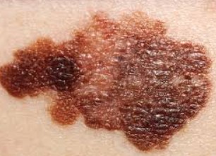
Asymmetric Melanoma — Leg
Uneven halves, irregular dark brown patches and a wider-than-6mm diameter. Classic asymmetry warning sign.
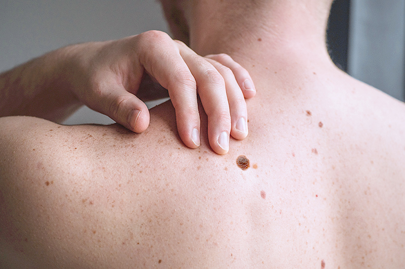
The 'Ugly Duckling' Sign — Back
One mole that looks distinctly different from the rest. Pattern-breaking lesions deserve a closer look.
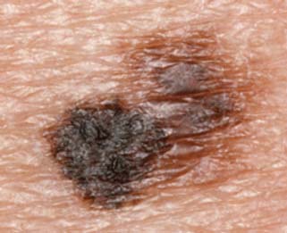
Irregular Border — Arm
Notched, scalloped edges and mixed black-brown colour. Borders that fade or blur into surrounding skin are a red flag.
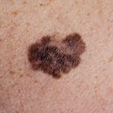
Multiple Colours — Back
Two or more shades of brown, black, red, white or blue within a single mole indicate concerning colour variation.
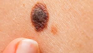
Large Diameter & Elevation — Leg
Diameter greater than 6 mm (pencil eraser) with raised, firm centre. Nodular melanomas grow downward and progress fast.
Does your mole look similar?
Get an instant AI risk assessment using the same ABCDE criteria dermatologists use.
When to seek medical help
Use the guidance below alongside — never instead of — your own judgement. AI screening cannot replace a clinical examination.
Arrange a routine assessment
- A new mole appearing after age 40
- A mole that looks distinctly different from your others
- You have a personal or family history of melanoma
Seek prompt advice (within ~2 weeks)
- Change in size, shape or colour over weeks/months
- Persistent itch, crust or new bleeding
- A pigmented streak under a fingernail or toenail
See NICE NG12 on suspected-cancer referral pathways.
Seek urgent help
- A rapidly growing pigmented or pink lesion
- An ulcerating, non-healing lesion
- New swollen lymph nodes near a suspicious mole
Seek medical advice even when an AI screening result appears low risk, if a lesion is changing, bleeding, painful or personally concerning. A low-risk result does not guarantee a mole is harmless.
What Does Basal Cell Carcinoma Look Like? 5 Real Examples
Basal cell carcinoma (BCC) is the most common skin cancer worldwide. It usually appears on sun-exposed areas as a pearly bump, a pink patch, or a sore that won't heal. Compare your spot to these dermatologist-referenced BCC photos.
Educational reference only — not a substitute for professional diagnosis.
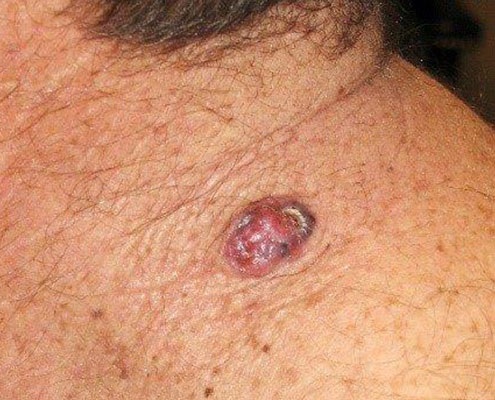
Pearly Nodular BCC — Shoulder
A shiny, pearly pink nodule with telangiectasia (tiny blood vessels) on chronically sun-exposed skin. The most classic BCC presentation.
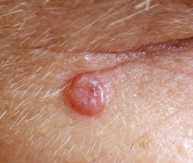
Periocular BCC — Eyelid Area
Translucent, dome-shaped papule near the eye. Eyelid and nose BCCs need prompt treatment to preserve surrounding tissue.
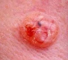
Ulcerated 'Rodent Ulcer' — Arm
Rolled, pearly border with a central crusted ulcer that bleeds and scabs repeatedly. Historically called a 'rodent ulcer'.
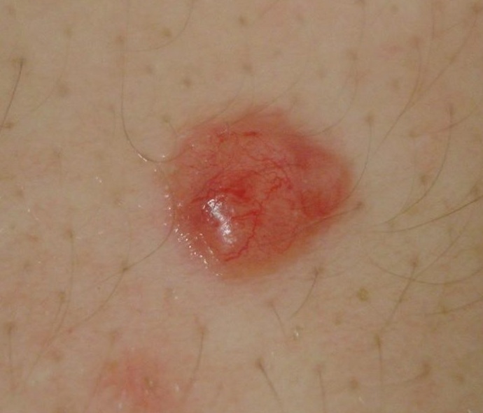
Superficial BCC — Back
Flat, pink-red scaly patch that may itch or bleed. Often misdiagnosed as eczema or psoriasis when it doesn't respond to creams.
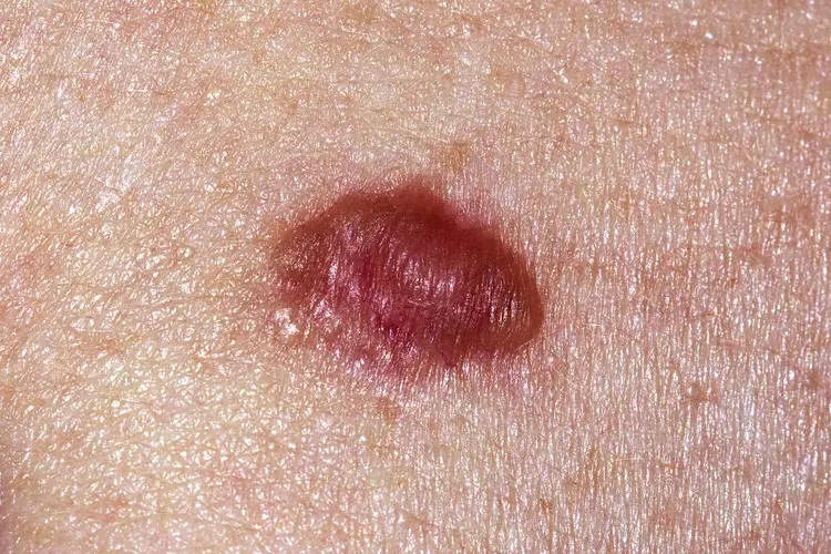
Pigmented BCC — Chest
Dark brown or purple BCC that can mimic melanoma. The pearly translucent edge is the key distinguishing feature.
Spot looks similar?
BCC is highly treatable when caught early. Get an AI risk indication in around 30 seconds.
What Does Squamous Cell Carcinoma Look Like? 5 Real Examples
Squamous cell carcinoma (SCC) is the second most common skin cancer. It typically appears as a scaly red patch, a thickened crusted bump, or a sore that won't heal. Compare your spot to these dermatologist-referenced SCC photos.
Educational reference only — not a substitute for professional diagnosis.
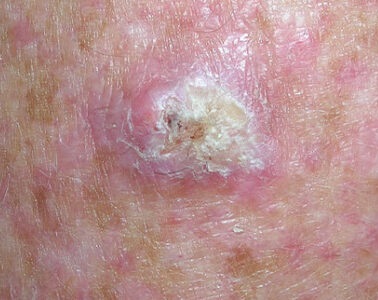
Hyperkeratotic SCC — Forearm
Thick, scaly white-yellow crust over a firm pink base on sun-damaged skin. The keratin build-up is a classic SCC sign.
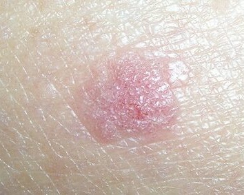
Scaly Pink Plaque — Chest
Raised pink plaque with a rough, scaly surface. Often mistaken for a wart or persistent dry patch that never heals.
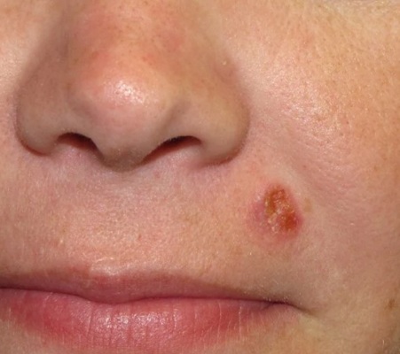
Facial SCC — Nose Area
Firm red-pink nodule on the central face. Facial SCC carries higher risk of spread and needs prompt dermatology review.
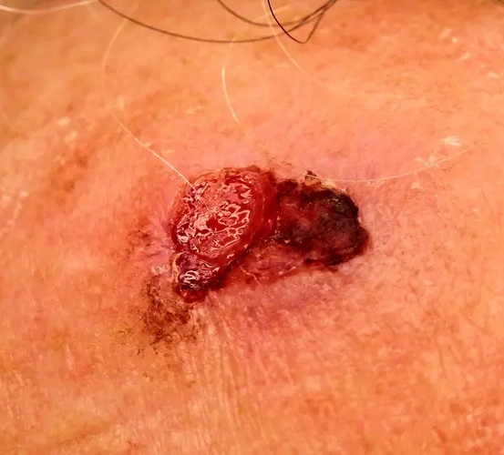
Ulcerated Advanced SCC
A raspberry-like raised lesion with bleeding and crusted ulceration. Advanced SCC needs urgent medical attention.
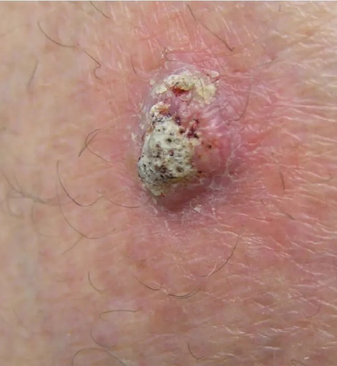
Crusted Non-Healing SCC — Back
Raised pink nodule with adherent thick scale that bleeds when knocked off. Any sore that won't heal in 4 weeks needs checking.
Sore that won't heal?
SCC is curable when caught early. Get an AI risk indication in around 30 seconds.
How to Take the Best Mole Photo
A good photo improves the quality of AI screening — but it does not guarantee an accurate result. Avoid filters and beauty modes, do not cover the area with makeup or cream, and retake the image if it is blurred or shadowed.
Good Lighting
Use bright, natural daylight. Avoid harsh shadows and flash glare.
Optional Scale
If you want a size reference, place a ruler next to the mole (preferred over a coin).
Sharp Focus
Hold the camera close enough for detail but still in focus. Clean the lens first.
Clean Background
Capture against clear skin. Avoid makeup, blood, or hair over the mole.
Understanding Your Results
Our AI provides a risk tier to help guide your next steps. These are screening results, not medical diagnoses.
The image did not show the same level of visible concern as higher-risk results. This is not a diagnosis and does not guarantee the mole is harmless. Continue monthly self-checks and see a clinician if the lesion changes, bleeds, itches or looks different from your other moles.
Some atypical visual features were detected. Consider a dermatologist review within a few weeks. Seek earlier assessment if the mole is changing, bleeding or painful — a clinician may need dermoscopy or biopsy for a definitive answer.
Multiple visually concerning features were identified. Please arrange a dermatologist assessment promptly. This is not a diagnosis; only a clinician can confirm with dermoscopy or biopsy.
Remember: These results indicate screening risk levels, not definitive diagnoses. Only a qualified dermatologist can diagnose skin conditions through professional examination and, if needed, biopsy.
Next Steps After Your Scan
Schedule a Dermatologist Visit
For medium or high risk results, book an appointment for dermoscopy and professional evaluation.
Track Changes Over Time
Use our chronic condition tracker to monitor your moles monthly and document any changes.
Regular Self-Exams
Check your entire body monthly—including scalp, between toes, and hard-to-see areas with help.
Why Regular Mole Checks Save Lives
Early detection matters
Cancer Research UK reports near-100% 5-year survival for Stage 1 melanoma, falling sharply at later stages. Source.
Peace of Mind
Regular monitoring reduces anxiety and helps you feel in control of your skin health.
Track Changes
Document mole evolution over time—the "E" in ABCDE is often the most important sign.
Why Choose Our AI Mole Checker?
ABCDE-based AI analysis
Analyses mole characteristics using the ABCDE method — 96.48% accuracy in internal benchmark testing.
Safe & Secure
Your images are processed securely and never stored without your permission
Instant Results
Get comprehensive mole analysis results within seconds
Easy to Use
Simply upload a clear photo of your mole for AI-supported screening
How our screening performs
The figure below describes agreement between our model and dermatologist labels — not a measure of clinical accuracy or diagnosis.
Clinical concordance (internal evaluation). View methodology.
Used by people in dozens of countries
Registered medical device module. See scope.
From Worried About a Mole to Reassured in 30 Seconds
Watch how a real user used ScanSkinAI to check a suspicious mole at home and decided on the right next step.
"I noticed a mole that looked different and didn't want to wait weeks for an appointment. I opened ScanSkinAI, took a photo, and within about half a minute I had a clear ABCDE-style summary and a sensible next step to book a review."
Personal user impression — AI screening is not a substitute for professional medical care.
- Instant ABCDE-style mole analysis from a single photo
- Clear risk indicators and next-step guidance
- Private, secure and available 24/7
Is your mole changing color or shape?
A mole changing color, growing, or developing an irregular shape is the single most important reason to keep an eye on it. Most changing moles are harmless — but change over time is what dermatologists ask about first, and it's what a one-off glance in the mirror can't measure. ScanSkinAI lets you check a suspicious mole free in your browser — no app to download, no signup needed to start — and then photograph the same spot over the following weeks to compare changes side by side.
Signs a mole should be checked
Get a mole looked at if it is changing in color (darkening, lightening, or developing multiple shades), changing shape or border, growing, itching, bleeding, or simply looks different from your other moles. If you've been asking yourself 'is this mole cancer?', the honest answer is that no photo tool can tell you — but a free AI mole check is a fast, private first step, and tracking the spot over time gives you and your doctor far better information than memory.
Why monitoring beats a one-off check
A single scan captures one moment. A mole changing shape or color reveals itself over weeks and months. Scanning the same spot regularly builds a visual timeline you can show a dermatologist — that's how changes get caught early.
Why one check is rarely enough
A single scan tells you about one spot, on one day. But skin changes are about patterns over time — a new mole appearing, a slow shift in shape, size or colour, or a patch that simply isn't healing. Monitoring the same spots side-by-side, week after week, surfaces the subtle changes a one-off check will always miss — and gives you a clear record to show a clinician if something needs a closer look.
(ScanSkinAI is a screening and monitoring tool, not a diagnosis. Always see a clinician for anything that is changing, bleeding, or worrying you.)
Unlimited scans · Track changes side-by-side · Cancel anytime
Related reading: track a mole over time · watching a mole for years?
Frequently Asked Questions
Everything you need to know about AI mole checking
In our internal evaluation, the model showed 96.48% accuracy in internal benchmark testing against dermatologist-labelled datasets, and 95.3% clinically acceptable concordance in post-market real-world use — screening metrics, not a confirmed diagnosis. The tool is designed for AI-supported screening to help you decide whether to seek professional review, and does not replace examination, dermoscopy or biopsy by a clinician.
Yes! Our basic mole checker is completely free with no hidden costs or credit card required. You can analyze multiple moles without any charges.
Our AI uses deep learning models trained on large public and licensed dermatology image datasets. From a single photo it can support visual assessment of Asymmetry, Border appearance and Colour variation. Diameter is only meaningful when you include a reliable scale (a ruler is preferred), and Evolution — change over time — can only be assessed by comparing the current image with an earlier image or documented history, so it is not measured from one photo.
Your photos are encrypted in transit and at rest. We do not sell your data or share images with third-party advertisers, and you control whether to save your results for tracking. The service is designed in line with UK GDPR data-protection principles — see our Privacy Policy for details.
If our AI indicates elevated risk, we recommend scheduling an appointment with a dermatologist for professional evaluation. Early detection significantly improves treatment outcomes for skin conditions.
Yes, you can check moles anywhere on your body. For hard-to-reach areas, ask someone to help take the photo. Ensure good lighting and a clear, focused image for the most accurate results.
We recommend monthly self-examinations and annual professional skin checks. Use our tool whenever you notice a new mole or changes in existing ones—size, shape, color, or texture changes warrant immediate attention.
Our AI can identify signs of melanoma, basal cell carcinoma, squamous cell carcinoma, benign nevi (normal moles), seborrheic keratosis, and other common skin lesions. It provides risk assessments and recommendations.
AI can screen for signs that may indicate melanoma based on visual characteristics like asymmetry, irregular borders, multiple colors, and size. However, a definitive diagnosis requires professional dermoscopy and potentially biopsy. Our tool helps identify moles that warrant medical attention.
Moles that warrant professional assessment often show ABCDE features: Asymmetry (uneven halves), irregular Borders, multiple Colors (brown, black, red, white, blue), a Diameter larger than about 6mm, and Evolution (changing over time). Evolution is judged by comparing photos or history — not from a single image. Any mole with these features should be evaluated by a dermatologist.
New moles appearing after age 30 should be monitored carefully, as most benign moles develop in childhood and adolescence. While not all new moles are dangerous, they warrant observation. Use our checker to screen new moles and consult a dermatologist if concerned.
Raised moles can be completely normal—many benign moles become elevated over time. However, a newly raised mole, or one that's changed from flat to raised, should be evaluated. Our AI can assess raised moles, but dermoscopy by a professional provides the most accurate evaluation.
Online mole checking with reputable AI tools like ours is safe as a screening method. Your data is encrypted and protected. However, it should complement, not replace, regular dermatologist visits. Never rely solely on AI for concerning symptoms—always seek professional evaluation.
AI mole checkers are most effective at detecting melanoma and pigmented lesions. They may be less accurate for non-pigmented skin cancers like amelanotic melanoma or some basal cell carcinomas. Comprehensive skin checks by a dermatologist remain essential.
Dermatologists use dermoscopy (specialized magnification), clinical experience, and can perform biopsies for definitive diagnosis. AI provides convenient preliminary screening to help you decide when to seek professional care. Both work best together—AI for regular monitoring, dermatologists for comprehensive evaluation.
Our AI is trained on diverse datasets including various skin tones. However, some skin cancers can present differently on darker skin. We continuously improve our algorithms for all skin types and recommend all users, regardless of skin tone, maintain regular professional dermatology check-ups.
For best results: use bright, even lighting (natural daylight is ideal), hold the camera close enough for sharp focus without losing detail (typically a hand's length away), clean the lens, avoid filters, beauty mode and heavy makeup, avoid flash glare, and capture the whole lesion with a little surrounding skin. If you want a size reference, a ruler is preferred over a coin. For tracking, use the same device, angle and lighting each time.
Analysis typically takes around 30 seconds depending on your device and connection. You'll receive a risk indication, ABCDE-style visual notes and next-step guidance shortly after uploading.
Our platform acts as an AI dermatologist to analyze your mole instantly. Upload a photo and get results in seconds. However, always consult a real dermatologist if concerns are identified—AI screening cannot replace professional medical evaluation.
No—AI analyzes moles to identify potential concerns, but it cannot remove them. For safe mole removal, you must consult a dermatologist who can assess whether removal is necessary and perform the procedure professionally. Never attempt to remove moles yourself.
Photograph it, check it with a free online mole checker, and monitor it over the following weeks. A mole changing color isn't automatically dangerous, but persistent change is a reason to see a doctor. ScanSkinAI is a screening and monitoring tool — it does not diagnose skin cancer or replace a dermatologist.
A suspicious mole check looks for asymmetry, irregular borders, uneven color, larger diameter, and evolution over time (the ABCDE signs). If any apply, or the mole itches, bleeds, or won't heal, arrange professional review. You can use ScanSkinAI's free mole checker in your browser as a first step and to track the spot between appointments.




Reviewed by our network of 17 dermatology clinicians
Meet the Clinical Review TeamEvidence & Safety
About Our AI Model
- • Trained on large public and licensed dermatology image datasets
- • Uses convolutional neural networks (CNNs)
- • Evaluated using standard ABCDE criteria
- • Regularly updated with new training data
- • 96.48% internal benchmark accuracy; 95.3% real-world dermatologist concordance
Bias & Limitations
- • Training data includes diverse skin tones, but biases may exist
- • Less effective for non-pigmented lesions
- • Photo quality significantly impacts results
- • Should not be used as sole diagnostic tool
- • Professional dermoscopy provides superior magnification
Your Privacy is Protected
- • Photos are encrypted in transit and at rest
- • Images are processed for analysis and deleted unless you choose to save them
- • We do not sell your data or share images with third-party advertisers
- • Designed in line with UK GDPR data-protection principles
Medical sources and further reading
Independent sources used to support guidance on this page. Links open in a new tab.
- NHS — Moles — symptoms and when to see a GP · accessed June 2026
- NHS — Melanoma skin cancer — overview · accessed June 2026
- NICE NG12 — Suspected cancer: recognition and referral · accessed June 2026
- Cancer Research UK — Melanoma survival statistics · accessed June 2026
- British Association of Dermatologists — Skin cancer patient information · accessed June 2026
- American Academy of Dermatology — ABCDEs of melanoma · accessed June 2026
- World Health Organization — Skin cancers — key facts · accessed June 2026
Learn More About Moles & Melanoma
ABCDE Rule for Melanoma
Complete guide to the 5 warning signs dermatologists use to identify potential melanoma.
The Ugly Duckling Sign
How to spot the mole that doesn't belong—a simple melanoma warning sign.
Mole vs Skin Tag vs Wart
Learn to tell the difference between common skin growths.
When to Worry About a Mole
12 red flags that should prompt a dermatologist visit.
How to Check a Mole
A simple 5-step self-check, what changes matter, and when to see a dermatologist.
New Mole as an Adult
What's normal, what's not, and when a new spot needs a professional check.
Educational Resources
Learn more about concerning mole symptoms and when to seek medical care
Choose the Right Tool for Your Concern
Based on what you're experiencing, one of these specialist tools may be more appropriate:
→ If You Suspect Melanoma
Deep-dive ABCDE analysis, melanoma stages, ugly duckling sign, and what to do next. Go here if your mole shows warning signs.
→ Skin Cancer Check Online
For sores that won't heal, crusty patches, pearly bumps, or other non-mole lesions that could be BCC or SCC.
→ Not Sure What It Is?
General AI skin analyzer for 80+ conditions including rashes, infections, and inflammatory conditions. Start here if unsure.
→ Track Changes Over Time
Monitor your moles monthly with photo comparison. Evolution is the most important warning sign.
Mole Guides
Explore expert-written guides to understand common symptoms, warning signs, and what to do next. These guides support your ScanSkinAI results and help you decide when to seek medical care.
ABCDE Rule for Melanoma
5 warning signs everyone should know
Ugly Duckling Sign
Find the mole that doesn't match
How to Check a Mole
Step-by-step self-exam guide
New Mole as an Adult
When new moles need attention
When to Worry About a Mole
Red flags you shouldn't ignore
Itchy Mole: When to Worry
Common causes vs warning signs
Bleeding Mole: What It Means
First aid and when to worry
Painful Mole: When to Worry
Is mole pain a sign of cancer?
Changing Mole Pictures
What visual changes mean
Melanoma vs Mole
Key differences explained
Early Detection Saves Lives
Don't wait. Get peace of mind with our free AI-powered mole checker today.
Important Medical Disclaimer
This AI tool provides preliminary screening and is not a substitute for professional medical diagnosis. Always consult a qualified dermatologist or healthcare provider for proper evaluation of suspicious moles or skin changes. Early professional diagnosis is crucial for effective treatment. If you have any concerning symptoms such as bleeding, rapid growth, or pain, seek medical attention immediately.
Mole Health Guides
When to Worry About a Mole
Learn the warning signs that mean a mole needs medical attention.
How to Check a Mole at Home
Step-by-step self-exam guide for monitoring your moles.
Melanoma vs Normal Mole
Visual comparison of melanoma warning signs versus benign moles.
Changes in Mole: What to Watch For
Photos and guidance on mole changes that need a dermatologist.