
Squamous Cell Carcinoma: Pictures, Symptoms & Treatment
Squamous cell carcinoma (SCC) develops from keratinocytes in the outer layers of the skin, usually on sun-damaged areas. It accounts for about 20% of skin cancers, and unlike basal cell carcinoma it carries a real, if small, risk of spreading.
Quick answer
Squamous cell carcinoma typically appears as a firm red nodule, a rough scaly patch, or a crusted sore that does not heal, on skin with long-term sun exposure. Cure rates exceed 95% with early surgical treatment, but 2–5% of cases metastasise, so any persistent scaly or ulcerated lesion should be examined by a clinician.
One free scan gives a first answer — the 3-month plan tracks a persistent sore or plaque over time.
Squamous cell carcinoma pictures
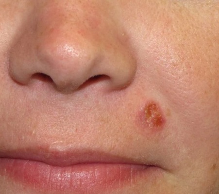
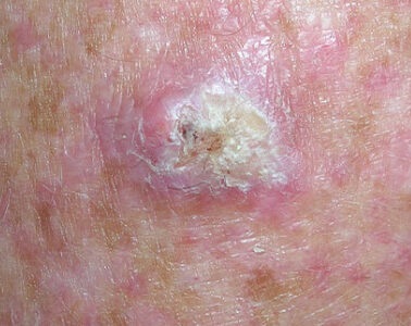
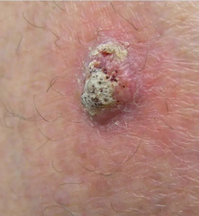
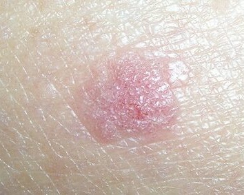
Compare with other types in our skin cancer pictures guide.
Early signs and symptoms
Common locations
- Face, ears and lips
- Bald scalp
- Neck and upper chest
- Forearms and backs of the hands
- Old scars, burns and chronic wounds
Warning signs
- Firm red nodule, sometimes tender
- Rough, scaly or crusted patch
- Sore that will not heal within four weeks
- Wart-like growth that bleeds
- New rough patch on the lip
- Any new lump arising in an old scar
SCC vs BCC vs actinic keratosis
| Condition | Typical look | Spread risk |
|---|---|---|
| Actinic keratosis | Rough, dry sandpaper-like patch; often easier to feel than see | Precancerous — 5–10% progress to SCC |
| Squamous cell carcinoma | Firm, scaly or crusted nodule; may ulcerate and bleed | 2–5% metastasise |
| Basal cell carcinoma | Pearly translucent bump with fine surface vessels | Under 0.1% metastasise |
Read the detailed comparison: squamous cell carcinoma vs actinic keratosis.
Risk factors and high-risk features
| Factor | Why it matters |
|---|---|
| Cumulative UV exposure | The dominant cause; outdoor workers are especially affected. |
| Existing actinic keratoses | Many SCCs arise from these precursor lesions. |
| Immunosuppression | Organ transplant recipients face up to 65× the risk and more aggressive tumours. |
| Location on lip or ear | These sites have a markedly higher metastatic risk. |
| Size over 2cm or deep invasion | Larger and deeper tumours recur and spread more often. |
| Chronic wounds and scars | Long-standing wounds can transform into SCC. |
| HPV infection | Associated with genital, periungual and some oral SCCs. |
Treatment options
Surgical excision
Standard care for most tumours, removing the lesion with a margin of healthy tissue and confirming clear edges under the microscope.
Mohs micrographic surgery
Highest cure rate, up to 99%. Used for high-risk tumours, recurrences, and lesions on the face, lips and ears where tissue preservation matters.
Curettage, radiotherapy and topical therapy
Curettage and electrodesiccation suit small, low-risk lesions. Radiotherapy is an option when surgery is not possible. Topical 5-FU or imiquimod may be used for SCC in situ.
Immunotherapy for advanced disease
Cemiplimab and pembrolizumab have substantially improved outcomes for locally advanced and metastatic SCC.
What else could it be? SCC look-alikes
Scaly, crusted or tender lesions are common and most are benign. These are the conditions most often mistaken for squamous cell carcinoma.
| Condition | How to tell it apart |
|---|---|
| Actinic keratosis | Rough sandpapery patch under 1cm that comes and goes with sun exposure. It is the precursor lesion — thickening, tenderness or bleeding suggests progression to SCC. |
| Seborrhoeic keratosis | Waxy, stuck-on plaque with a crumbly surface. Not tender, does not ulcerate. |
| Wart (verruca) | Rough papule with pinpoint black dots (thrombosed capillaries), often multiple, and it interrupts skin lines. |
| Basal cell carcinoma | Pearly and translucent with fine surface vessels rather than thick keratin scale. |
| Keratoacanthoma | Dome-shaped nodule with a central keratin plug that erupts within weeks. Clinically indistinguishable from SCC — it is excised and examined. |
| Chronic eczema or psoriasis plaque | Symmetrical, itchy and responsive to topical steroid. A single fixed, growing, tender plaque is not eczema. |
Scaly patch or sore that will not heal?
Photograph it and the AI skin check screens for the features associated with squamous cell carcinoma — thick keratin scale, ulceration, rapid growth — with a suggested next step.
When to see a dermatologist
Get checked promptly for
- Any sore that has not healed in four weeks
- A scaly patch that thickens, hardens or becomes tender
- A growth that bleeds repeatedly or crusts over
- A new lesion on the lip or ear
- A rapidly enlarging lump, especially if you are immunosuppressed
After an SCC diagnosis, 30–50% of people develop another within five years, so ongoing skin surveillance is essential.
Frequently asked questions
What does squamous cell carcinoma look like?
SCC usually appears as a firm red nodule, a rough scaly patch, a crusted sore that will not heal, or a wart-like growth that bleeds. It is most common on sun-exposed skin: face, ears, lips, scalp, neck, forearms and backs of the hands.
How serious is squamous cell carcinoma?
Most SCCs are highly curable, with cure rates above 95% when treated early. Unlike basal cell carcinoma, SCC can spread — around 2–5% metastasise to lymph nodes or beyond — so it should not be left alone.
How fast does squamous cell carcinoma grow?
SCC grows faster than basal cell carcinoma, typically becoming noticeable over weeks to months. A lesion that enlarges quickly, becomes tender, or ulcerates should be assessed without delay.
Does actinic keratosis turn into squamous cell carcinoma?
It can. Actinic keratosis is a precancerous lesion and an estimated 5–10% of untreated lesions progress to SCC over a decade. Treating actinic keratoses and protecting the surrounding sun-damaged field is the main way to reduce that risk.
What makes an SCC high risk?
High-risk features include size over 2cm, poor differentiation, depth beyond the fat layer, perineural invasion, location on the lip or ear, arising in a scar or chronic wound, recurrence after treatment, and immunosuppression. These cases usually need Mohs surgery and close follow-up.
What is the treatment for squamous cell carcinoma?
Surgical excision with clear margins is standard. Mohs micrographic surgery is preferred for high-risk or cosmetically sensitive sites. Superficial SCC in situ may be treated topically or with curettage. Advanced or metastatic SCC is treated with immunotherapy such as cemiplimab or pembrolizumab.
Unsure about a scaly patch or sore?
Upload a clear photo for a free AI skin check covering SCC, BCC, melanoma and precancerous changes.
Free skin cancer check online