
TL;DR: Key Takeaways
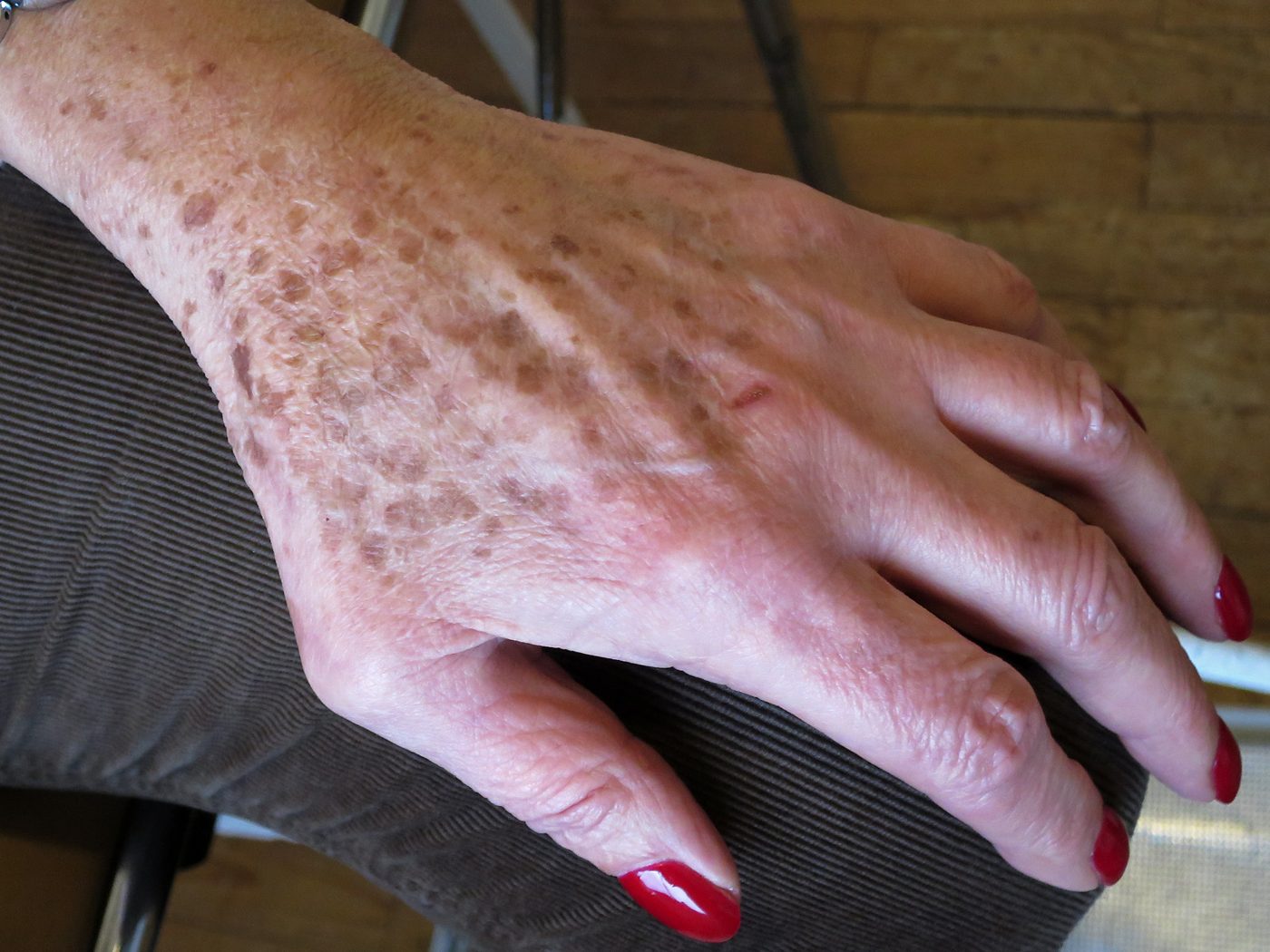
- Solar lentigines (age spots) = flat, uniform, stable, harmless
- Lentigo maligna melanoma = asymmetric, multi-shaded, slowly enlarging — cancer
- Most common on chronically sun-exposed skin after age 60, especially face
- Lentigo maligna grows over years — easy to miss without photo comparison
- Any age spot that darkens unevenly, enlarges, or develops a raised area → see a dermatologist
Part of the Sun Damage vs Skin Cancer hub.
At a Glance
| Feature | Solar Lentigo | Lentigo Maligna Melanoma |
|---|---|---|
| Colour | Uniform tan to medium brown | Variegated — tan, dark brown, black, occasionally blue or pink |
| Border | Smooth, well-defined | Irregular, notched, fading into normal skin |
| Symmetry | Symmetric | Asymmetric |
| Size | 2–20 mm, stable | Often >6 mm, slowly enlarging over years |
| Surface | Completely flat and smooth | Flat early; can develop a raised nodule late (invasive stage) |
| Age of onset | From 40s onward | Usually after 60 |
| Change over time | Stable for years | Slow enlargement and darkening over 2–10+ years |
| Risk level | None | In-situ melanoma; becomes invasive if untreated |
How a Solar Lentigo Looks
- Flat brown patch, smooth to the touch
- Uniform single shade of tan or brown
- Round or oval with clean borders
- Usually one of many similar spots in the same area
- Stable for years — looks identical in photos from 5 years ago
How Lentigo Maligna Looks
- Asymmetric flat patch on chronically sun-exposed skin (face, neck, forearms)
- Multiple shades of brown to black within the same lesion
- Irregular notched borders that fade into surrounding skin
- Slowly enlarging — often noticed when comparing to old photos
- Late stage: a raised dark nodule develops within the flat patch (invasive melanoma)
Not sure which one you have?
Upload a photo for an instant AI screen — covers melanoma, BCC, SCC and 80+ conditions.
The 5 Dermatologist Clues
- Asymmetry — fold the spot in half mentally; lentigo maligna rarely matches
- Colour variegation — three or more shades in one lesion is a red flag
- Border irregularity — fading or notched borders favour melanoma
- Diameter >6 mm — not diagnostic alone, but raises concern in combination
- Evolution — slow growth visible only by comparing old photos is the classic lentigo maligna pattern
Red-Flag Checklist
- An age spot that has darkened or enlarged compared to 1–2 years ago
- More than two colours within a single 'age spot'
- A flat dark patch on the face or neck that's slowly growing
- Any age spot that develops a raised area within it
- An 'age spot' that looks distinctly different from your others
If any of these apply, book a dermatologist within 2–4 weeks.
Scan this mole free — no app needed
Upload one photo. Get an AI screening for melanoma, BCC and 80+ skin conditions in seconds. Works in Safari and Chrome.
Frequently Asked Questions
A solar lentigo (age spot) is a flat, uniformly pigmented, stable benign lesion caused by chronic sun exposure. Melanoma — especially lentigo maligna melanoma, the type that arises on sun-damaged skin — is asymmetric, has uneven colour, slowly enlarges, and is a potentially life-threatening cancer.
A solar lentigo itself does not transform into melanoma. However, lentigo maligna melanoma develops on the same chronically sun-damaged skin and can mimic an age spot in its early years — which is why any age spot that changes deserves evaluation.
Very slowly — often visible enlargement over years, not months. This makes it easy to miss. The key signs are uneven darkening, irregular borders, and gradual enlargement of a 'spot' that used to look like all the others.
Fair-skinned adults over 60 with significant lifetime sun exposure, particularly on the face, neck, scalp, and forearms. People with many solar lentigines are at higher risk.
Yes — definitive diagnosis requires a skin biopsy. Dermoscopy and AI screening can flag suspicious lesions, but only histopathology confirms melanoma.