
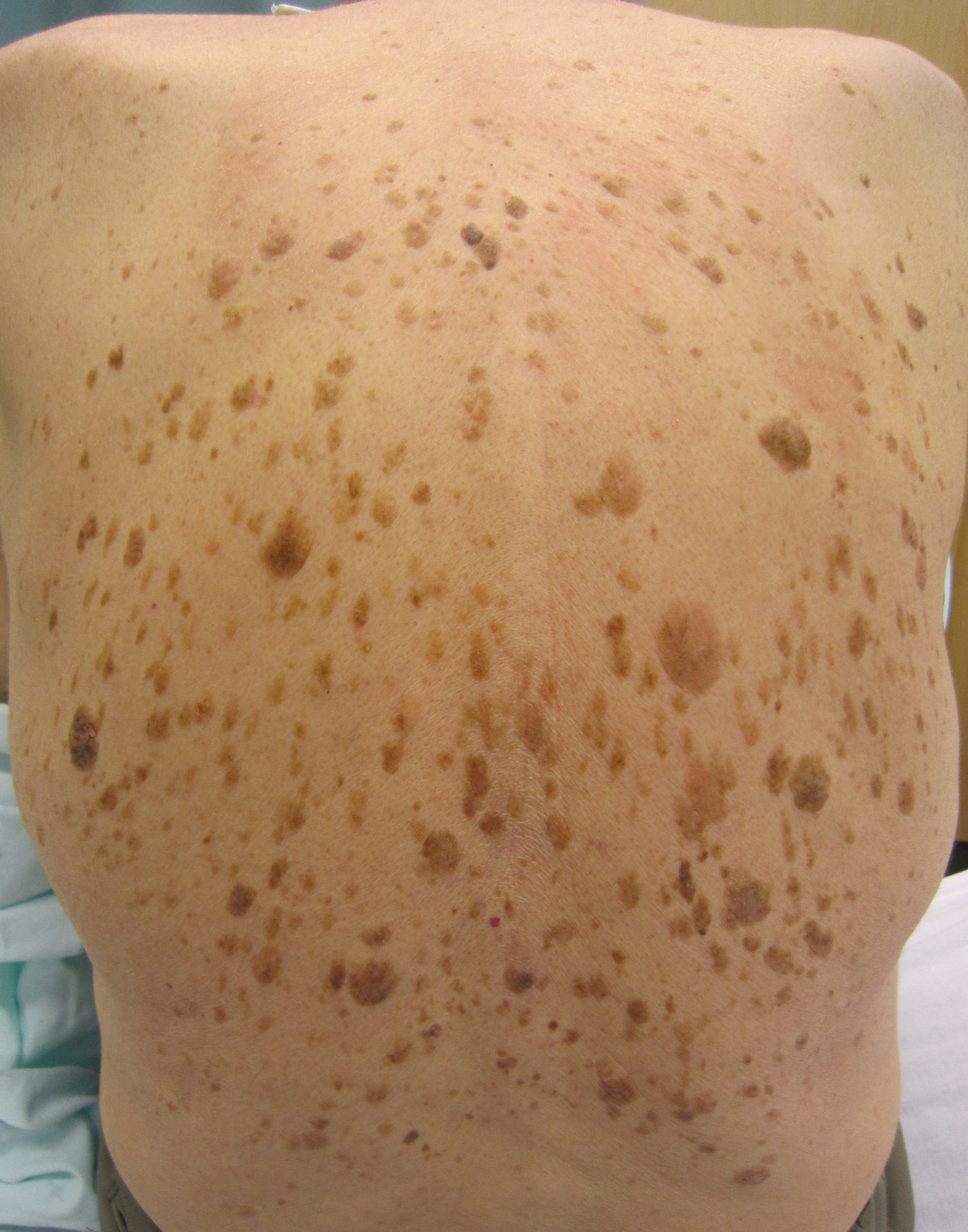
TL;DR: Key Takeaways
- Seborrheic keratoses are benign — they never become cancer
- But they're the #1 melanoma mimic — pigmented SKs can look identical to melanoma
- Key tell: waxy, 'stuck-on' surface that looks pasted onto the skin
- SKs are stable for years; melanoma changes over months
- When in doubt, photograph and use AI screening or see a dermatologist
Part of the Sun Damage vs Skin Cancer hub.
At a Glance
| Feature | Seborrheic Keratosis | Melanoma |
|---|---|---|
| Surface | Waxy, "stuck-on", often crumbly | Smooth or slightly raised, integrated with skin |
| Colour | Tan, brown, or black — usually one shade per lesion | Multiple shades within one lesion (brown, black, pink, blue) |
| Border | Well-defined, often round | Notched, scalloped, or irregular |
| Texture if scraped | Scale lifts easily (don't actually do this) | Does not flake off — integrated with skin |
| Number | Usually multiple, often dozens | Almost always a single 'ugly duckling' lesion |
| Location | Trunk, face, anywhere | Anywhere — back, legs, and sun-exposed sites most common |
| Change over time | Stable for years | Changes over weeks to months |
| Danger | None | Cancer — survival depends on early detection |
How a Seborrheic Keratosis Looks
- • 'Stuck-on' waxy surface
- • Uniform tan/brown/black
- • Sharp round border
- • Often multiple
- • Stable for years
- • Asymmetrical shape
- • Mixed colours in one lesion
- • Notched / fading border
- • Usually solitary
- • Changing over months
Illustrative diagrams. For verified clinical photos, see the skin diseases A–Z.
- Waxy, 'stuck-on' appearance — as if pasted onto the skin
- Surface often has small horn cysts or pinpoint dots visible up close
- Colour ranges from skin-tone to tan, brown, or black
- Usually round or oval with a sharp border
- Often part of a 'family' — most adults over 50 have several
How Melanoma Looks
- Asymmetric — one half does not match the other
- Multiple colours within the same lesion
- Borders that fade or notch into surrounding skin
- Visibly changing over months
- Stands out as the 'odd one' compared to your other moles or SKs
Not sure which one you have?
Upload a photo for an instant AI screen across melanoma, BCC, SCC and 80+ conditions.
The 5 Visual Clues Dermatologists Use
- 'Stuck-on' surface — strongest single feature of SK
- Horn cysts — tiny white or yellow dots visible on the SK surface
- Multiple similar lesions nearby — suggests SK, not melanoma
- Asymmetric multi-coloured appearance — suggests melanoma
- Recent change — strongest single feature of melanoma
Red-Flag Checklist
- Sudden eruption of many new SKs ('Leser-Trélat sign') — see a doctor; rarely linked to internal cancer
- A long-standing SK that develops new colours, bleeds, or grows — rule out adjacent melanoma
- A single 'odd duckling' lesion with multi-colour appearance — needs evaluation within 2–4 weeks
- Itching, bleeding or pain in any pigmented lesion — see a dermatologist
Scan this mole free — no app needed
Upload one photo. Get an AI screening for melanoma, BCC and 80+ skin conditions in seconds. Works in Safari and Chrome.
Frequently Asked Questions
No — seborrheic keratoses are completely benign and never become cancerous. The concern is that they can look strikingly like melanoma, so misidentification (in either direction) is the actual risk.
Seborrheic keratoses have a waxy, 'stuck-on' appearance, are stable over years, and usually have a uniform crusty surface. Melanoma is typically asymmetric, has multiple colours within one lesion, and changes over months. When in doubt, get a dermatologist or AI screen.
No. Seborrheic keratoses do not transform into melanoma. But melanoma can rarely develop right next to or within a seborrheic keratosis — which is why any new colour, bleeding, or change in a long-standing SK still deserves evaluation.
Because pigmented seborrheic keratoses can be black, irregular-bordered, and asymmetric — checking every box on the ABCDE melanoma rule. The 'stuck-on' surface texture is the giveaway, but it requires close inspection.
Only if they're symptomatic (irritated, catching on clothes, bleeding from friction) or for cosmetic reasons. Removal options include cryotherapy, curettage, and electrocautery.