
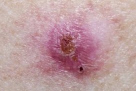
What is Basal Cell Carcinoma?
Basal cell carcinoma (BCC) is a type of skin cancer that begins in the basal cells—the cells at the bottom of the epidermis that produce new skin cells. It's the most common cancer in humans, with millions of cases diagnosed each year.
The good news: BCC is highly treatable, especially when caught early. It rarely spreads to other parts of the body, but if left untreated, it can grow deep into tissue, causing significant local damage.
Types of Basal Cell Carcinoma
Nodular BCC
Shiny, dome-shaped bump, often pearly or pink. May have visible blood vessels. Most common type.
Superficial BCC
Flat, reddish, scaly patch that may resemble eczema or psoriasis. Often on trunk.
Morpheaform BCC
Waxy, scar-like area with poorly defined borders. Can be skin-coloured or white. An infiltrative subtype that may have less clearly defined borders.
Pigmented BCC
Dark brown or black coloring within the lesion. Can be mistaken for melanoma.
What Does Basal Cell Carcinoma on the Face Look Like?
Basal cell carcinoma on face is common because facial skin receives high cumulative UV exposure. Lesions may appear on the nose, eyelids and inner eye area, ears, cheeks, forehead, lips or nearby skin, and the scalp in areas with thinning hair.
Possible appearances include a small shiny or pearly bump, visible fine blood vessels (telangiectasia), a sore that bleeds, crusts, heals and returns, a flat scaly patch, a waxy or scar-like area, or a brown or pigmented growth that may be mistaken for a mole. BCC can also look different across skin tones — it may appear brown, black, pigmented or similar in colour to the surrounding skin.
Because BCC on the face is close to the eyes, nose and lips, even small lesions are worth reviewing early — a clinician may need a biopsy to confirm what the growth is.
Warning Signs of BCC
See a doctor if you notice any of these signs:
BCC can look different across skin tones and may appear brown, black, pigmented or similar in colour to the surrounding skin.
Common Locations for BCC
BCC commonly appears on sun-exposed areas, although it can develop elsewhere:
Frequently Asked Questions
Early BCC often appears as a small, shiny or pearly bump (often on the face), a flat pinkish, reddish or brown patch, a waxy or scar-like area, or a small growth with raised edges. Tiny blood vessels may be visible on the surface, and appearance can vary across different skin tones.
BCC is generally less aggressive than other skin cancers because it rarely spreads to other parts of the body. However, if left untreated it can grow into surrounding skin, muscle, and bone, causing significant local damage. Early assessment and treatment are important.
BCC commonly appears on sun-exposed areas: face (especially nose, eyelids, lips), ears, scalp (in areas with thinning hair), neck and shoulders. BCC can also occur outside sun-exposed areas, so any new or changing lesion is worth checking.
BCC usually grows slowly over months to years, and some lesions remain stable for long periods. Growth rate varies between subtypes, so even a slow-growing lesion should be reviewed rather than ignored.
BCC does not reliably go away on its own and typically requires medical treatment. Without treatment it can continue to grow slowly and cause tissue damage. Home remedies do not treat BCC.
The primary risk factor is cumulative UV exposure from sunlight or tanning beds. Other contributors include fair skin, light eyes, history of sunburns, a weakened immune system, radiation exposure, and certain genetic syndromes.
A clinician may need a biopsy to confirm BCC. Treatment depends on the lesion's type, size and location, and can include surgical excision, Mohs surgery (often used on the face), curettage and electrodesiccation, cryotherapy, topical medications for superficial BCC, or radiation therapy.
BCC very rarely spreads to distant organs. However, it can extend locally into surrounding tissue, bone, and nerves if left untreated for years, which is why early assessment matters.
A BCC may appear as a sore that bleeds easily, heals partially then reopens, crusts or oozes, has a central depression or crater, and has not healed after about four weeks. A sore that repeatedly heals and returns is a classic BCC pattern.
No. Many benign lesions can appear shiny, including sebaceous hyperplasia, molluscum, and some moles. However, a new, persistent or slowly growing shiny bump — especially on sun-exposed skin — is worth having checked. A biopsy may be needed to confirm BCC.
BCC is more common after age 50 but can occur in younger adults, especially those with significant sun exposure, fair skin, or tanning bed use. BCC can also look different across skin tones.
Prevention includes broad-spectrum sunscreen, protective clothing and hats, avoiding peak UV hours, avoiding tanning beds, seeking shade, and regular skin self-exams. Report any new, changing or non-healing lesion to a clinician.
Screen a Spot You're Worried About
Our AI-supported tool analyses visible skin features associated with common skin concerns. The result is not a diagnosis.
Screen This SpotMedical Disclaimer
This article is educational only. Only a dermatologist can diagnose BCC. If you notice suspicious growths, seek professional evaluation promptly.