
Pyogenic Granuloma: Causes, Treatment & How to Tell It Apart from Amelanotic Melanoma
Medically reviewed by Dr. Celina Kazumi Iwasa, MD, Dermatology · Last updated June 2026
A rapidly growing, bright red or purplish bump made of overgrown blood vessels. Despite the name, it's neither infectious (pyogenic) nor a true granuloma — it's a benign vascular lesion that bleeds easily, often after minor trauma, and is common in children, pregnant people, and on the fingers, lips, and gums.
Concerned this might be pyogenic granuloma? Scan it with our AI skin checker or, for moles and pigmented spots, our AI mole checker. Free, no signup required.
Quick Answer
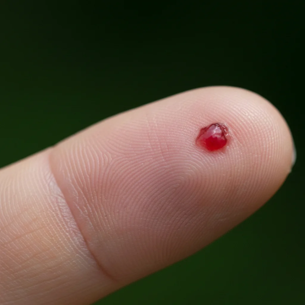
A pyogenic granuloma is a fast-growing, bright red or purplish bump made of overgrown blood vessels. Despite its name, it is not an infection. These common, benign growths typically appear over a few weeks and bleed very easily, often after a minor scrape. They most commonly show up on the fingers, face, lips, or gums, particularly in children and pregnant people. Because they bleed so easily and can sometimes look like serious skin cancers, it is important to have a doctor examine and remove them.
Clinical Context
Pyogenic granuloma (lobular capillary hemangioma) is a common acquired benign vascular proliferation. Lesions typically grow rapidly over weeks to a few centimetres, ulcerate, and bleed profusely with minor trauma. They most often arise on the head, neck, fingers, and oral mucosa. Pregnancy-associated lesions ('granuloma gravidarum') frequently appear on the gums in the second trimester and may regress postpartum. The main clinical concern is misdiagnosis: amelanotic melanoma, Kaposi sarcoma, and basal cell carcinoma can mimic pyogenic granuloma, so histological confirmation after excision is standard practice.
Symptoms
- Bright red, purple, or brownish bump
- Rapid growth over days to weeks
- Smooth, glistening or ulcerated surface
- Bleeds easily — often profusely — with minor trauma
- Usually 5–10 mm but can reach 2 cm
- Often has a collarette of scale at the base
- Most common on fingers, lips, gums, scalp, and face
Severity & Progression
Causes & Risk Factors
- Minor skin trauma (cuts, burns, insect bites)
- Hormonal changes — pregnancy and oral contraceptives
- Certain medications (oral retinoids, indinavir, EGFR inhibitors)
- Underlying vascular malformation (less common)
- Idiopathic — no identifiable trigger in many cases
How Pyogenic Granuloma Differs from Similar Conditions
Several conditions can look similar. Here's how to tell them apart — though a healthcare professional can provide a definitive diagnosis.
| Condition | Key Difference from Pyogenic Granuloma |
|---|---|
| Amelanotic Melanoma | Grows over months rather than weeks, often has subtle pink, red, or skin-coloured asymmetry, less likely to bleed spontaneously at small sizes, and arises in adults. Biopsy is mandatory whenever pyogenic granuloma is suspected in an adult. |
| Cherry Angioma | Small (1–5 mm), stable for years, dome-shaped, rarely bleeds unless traumatised. Pyogenic granuloma is larger, grows rapidly, and bleeds at the slightest touch. |
| Basal Cell Carcinoma (nodular) | Pearly translucent appearance with visible telangiectasias and a rolled border; grows over months to years, not weeks. May ulcerate but does not bleed as readily as pyogenic granuloma. |
| Kaposi Sarcoma | Usually multiple purple-brown patches and plaques, typically on the legs or oral mucosa, and associated with HHV-8 and immunosuppression. Pyogenic granuloma is almost always solitary. |
Treatment & Management
Because pyogenic granuloma rarely regresses on its own and bleeds repeatedly, complete removal is usually recommended. Shave excision with curettage and electrocautery of the base is the most common in-office procedure and gives a histology specimen to rule out melanoma. Full-thickness excision is preferred for larger or recurrent lesions because it has the lowest recurrence rate (<5%). Pulsed dye laser and topical timolol or imiquimod are useful for small lesions in children where surgery is poorly tolerated. Pregnancy-associated oral lesions are often left alone if asymptomatic and frequently shrink after delivery.
- Shave excision with curettage and cautery (first-line)
- Full-thickness excision for large or recurrent lesions
- Pulsed dye laser (small lesions, children)
- Topical timolol 0.5% gel (small lesions)
- Topical imiquimod 5%
- Silver nitrate cautery (limited use)
- Observation only for oral pregnancy lesions
Red Flags & Complications
Seek medical attention if you experience any of the following:
- Recurrent heavy bleeding
- Iron-deficiency anemia from repeated bleeding (rare)
- Recurrence after incomplete removal (15–40%)
- Satellite lesion formation
- Cosmetic scarring
- Misdiagnosis of amelanotic melanoma if not biopsied
Pyogenic Granuloma Across Skin Types and Hair Types
Pyogenic granuloma looks bright red to purple on lighter skin (Fitzpatrick I–III). On medium and darker skin (Fitzpatrick IV–VI) the lesion appears deeper red-brown or violaceous and may be harder to distinguish from a melanocytic lesion by colour alone — texture, the collarette of scale, and the history of rapid growth become the most reliable clues.
Self-Care Tips
- Apply firm pressure with clean gauze for 10–15 minutes to stop bleeding
- Cover with a non-stick dressing between episodes
- Avoid picking or trying to remove the lesion at home
- Photograph weekly to document growth before your appointment
- Seek same-day care for bleeding that does not stop with pressure
When to See a Doctor
See a clinician within 1–2 weeks for any new, fast-growing red bump that bleeds easily — especially if it's larger than 5 mm, arises without trauma, or appears in an adult. Histology after removal is the standard of care to rule out amelanotic melanoma.
Frequently Asked Questions
What does a pyogenic granuloma usually look like?
It typically looks like a small, bright red, shiny bump that grows very fast over just a few days or weeks. It might look a bit like a raspberry and often has a small ring of scaly skin at its base. The most noticeable symptom is that it bleeds heavily and repeatedly, even with the slightest bump or friction.
Why did I get this bleeding red bump?
The exact cause isn't always known, but they frequently appear after a minor skin injury, like a scratch, bug bite, or burn. Hormonal changes are another major trigger, which is why they are very common on the gums of pregnant women. Certain medications, like acne pills and some chemotherapy drugs, can also cause these overgrown blood vessels to develop.
Could this red bump be amelanotic melanoma?
Yes, it is possible for amelanotic melanoma—a type of skin cancer that lacks dark brown or black pigment—to look almost exactly like a pyogenic granuloma. Because both can be fast-growing, reddish bumps that bleed, doctors take these spots very seriously. A doctor will usually remove the bump and send it to a lab for testing just to be completely sure it is benign.
How do doctors treat a pyogenic granuloma?
Because these bumps rarely go away on their own and bleed so much, complete removal is the standard treatment. A doctor will typically perform a shave excision, scraping the bump off and then using heat to seal the base. For larger or recurring bumps, they might cut it out entirely and use stitches. Sometimes, lasers or special topical creams are used.
Will the bump come back after it is removed?
When treated properly by a medical professional, the prognosis is excellent. However, if the entire base of the blood vessel isn't completely destroyed during removal, a pyogenic granuloma can grow back. Full surgical removal offers the lowest chance of it returning, at less than five percent. If it does come back, your doctor may suggest a different removal method.
When should I see a doctor about a bleeding bump?
You should see a doctor promptly if you develop any new, fast-growing bump that bleeds easily, oozes, or does not heal. Because a pyogenic granuloma shares visual characteristics with certain serious skin cancers, an accurate medical evaluation is essential. Do not try to pop or cut off the bump at home, as it will likely bleed profusely and regrow.
Is a pyogenic granuloma contagious?
No, pyogenic granulomas are completely harmless to others and are not contagious. The name is quite misleading; 'pyogenic' implies it is caused by a bacterial infection that produces pus, but it is actually just an overgrowth of small blood vessels. Because they are tied to minor injuries or hormones, there is no surefire way to prevent them.
How can ScanSkinAI help me check a fast-growing red bump?
ScanSkinAI can help you monitor changes in your skin and provide an initial analysis of your spot. By analyzing a photo, the AI acts as an educational screening aid to compare your bump's features against its database. However, AI cannot diagnose a pyogenic granuloma or rule out amelanotic melanoma. Always consult a healthcare professional for a medical diagnosis and treatment plan.
Related Symptoms
Explore symptom guides that may help you understand this condition better:
Further Reading
Related Conditions
Medical References
Information on this page is sourced from and verified against reputable medical resources:
Concerned About Your Skin?
Upload a photo and get instant AI-powered analysis across 80+ skin conditions. Track your condition over time with photo timelines, or use our dedicated AI mole checker for moles and pigmented lesions.
Medical Disclaimer: This information is for educational purposes only and is not intended as medical advice. The content on this page should not be used to diagnose or treat any health problem. Always consult with a qualified healthcare professional for proper medical evaluation, diagnosis, and treatment of your condition.