
Actinic Keratosis Pictures: What It Really Looks Like
Actinic (solar) keratosis is the most common precancer — a rough patch of sun-damaged skin that can progress to squamous cell carcinoma if ignored. It is usually easier to feel than to see, which is why photos alone never tell the whole story.
Medically reviewed by Dr. Celina Kazumi Iwasa, MD · Last reviewed August 2026
Quick answer
Actinic keratosis looks like a small rough, scaly patch — skin-toned, pink, red or brown — on sun-exposed skin such as the face, ears, scalp and backs of hands. The defining feature is texture: it feels like sandpaper even when it is barely visible. Any rough patch that persists for more than a few weeks, thickens, bleeds or grows a hard scale should be checked by a doctor, because a proportion of untreated lesions become squamous cell carcinoma.
One free scan gives a first answer — the 3-month plan tracks rough, sun-damaged patches over time.
Clinical picture
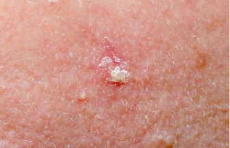
More early-stage images: actinic keratosis pictures, early stage · Full guide: actinic keratosis symptoms, causes and treatment
The four patterns to recognise
Feels like sandpaper
The single most reliable early clue: a rough, gritty patch you can feel with a fingertip before you can clearly see it.
Sun-exposed sites
Face (cheeks, nose, forehead), ears, bald scalp, neck, backs of hands and forearms — where a lifetime of UV lands.
Colour varies
Skin-toned, pink, red or brown. On darker skin tones patches may look grey or slightly darker than surrounding skin.
Scale or horn
More advanced lesions build a hard scale, crust or even a small cutaneous horn — a sign to be seen promptly.
What it is commonly confused with
| Lookalike | How it differs |
|---|---|
| Sun spot (lentigo) | Flat and smooth with no scale — purely a colour change. |
| Seborrhoeic keratosis | Waxy, raised, 'stuck-on' appearance; benign but can look alarming. |
| Squamous cell carcinoma | Thicker, firmer, faster-growing; may ulcerate or bleed. This is what AK can progress to. |
| Eczema / dry skin | Improves with moisturiser; AK does not, and stays in sun-exposed sites. |
See a doctor promptly if
- A rough patch thickens, hardens or grows a horn-like scale
- The patch bleeds, ulcerates or becomes tender
- A lesion grows noticeably over weeks
- You have many patches across sun-damaged skin (field change)
Frequently asked questions
What does actinic keratosis look like in pictures?
Actinic keratosis typically appears as a small, rough, scaly patch — often described as feeling like sandpaper. Colours range from skin-toned and pink to red or brown. Patches are usually easier to feel than to see and appear on sun-exposed areas: face, ears, scalp, backs of hands and forearms.
How can you tell actinic keratosis apart from a normal sun spot?
A sun spot (lentigo) is flat and smooth; actinic keratosis has a rough, gritty or scaly surface you can feel with a fingertip, sometimes with a hard scale or horn. AK patches may be tender, itch, or sting. Any rough patch that persists for more than a few weeks on sun-exposed skin should be checked.
Can actinic keratosis turn into skin cancer?
Yes. Actinic keratosis is precancerous: a proportion of untreated lesions progress to squamous cell carcinoma. Most never do, but because it is impossible to predict which will, dermatologists treat visible lesions — commonly with cryotherapy or prescription field creams.
What does early actinic keratosis look like?
Early lesions are often tiny, faint and feel like a dry rough spot that does not moisturise away. They may look pink on lighter skin or grey-brown on darker skin. Catching them at this stage makes treatment simple and prevents progression.
When should I see a doctor about a rough patch?
See a doctor if a rough, scaly patch persists beyond 3–4 weeks, grows, thickens, bleeds, becomes tender, or develops a hard horn. A rapidly growing or ulcerating lesion on sun-damaged skin needs prompt review to rule out squamous cell carcinoma.
This page is for education and screening support only and does not replace medical advice. ScanSkinAI is not a diagnostic device — always consult a qualified clinician about any changing skin lesion.