
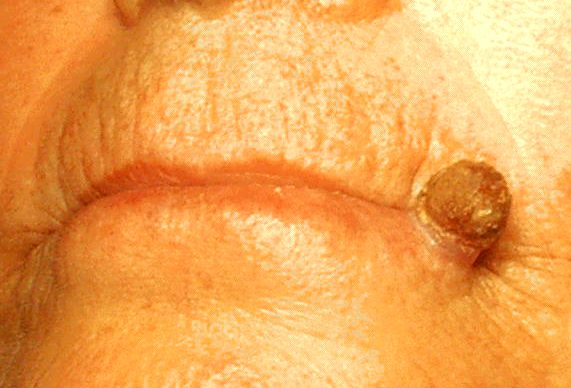
TL;DR: Key Takeaways
- Early AK = small, rough, scaly patch on chronically sun-exposed skin
- Often easier to feel (sandpaper texture) than to see
- Pink, red, or skin-coloured base with a dry scale on top
- Face, scalp (bald men), ears, backs of hands, forearms are typical sites
- Precancerous — 5–10% progress to SCC over years; all are treatable
Part of the Sun Damage vs Skin Cancer hub.
What Early Actinic Keratosis Looks Like
- Size — usually 2 mm to 6 mm in earliest stage, can grow to 1 cm or more
- Texture — rough and scaly, often more obvious to touch than to look at
- Colour — pink, red, light brown, or skin-coloured base; scale on top is usually whitish-yellow
- Shape — irregular, often poorly defined edges
- Number — usually multiple AKs cluster together; isolated single AKs are less common
Where AKs Appear on the Body
AKs only develop on chronically sun-exposed skin. The classic sites:
- Face — forehead, temples, cheeks, nose, lower lip (actinic cheilitis)
- Scalp — particularly common in bald or thin-haired men
- Ears — top rim and earlobes in men; behind the ears in women
- Backs of hands and forearms
- Lower legs in women who wore skirts and dresses
- Upper chest and shoulders with chronic outdoor exposure
Compare your spot with AI
Upload a photo for an instant AI screen across AK, SCC and 80+ conditions — no signup.
AK vs the 4 Common Lookalikes
| Condition | Key Distinguishing Feature |
|---|---|
| Sun spot (solar lentigo) | Completely flat and smooth — no rough scale |
| Seborrheic keratosis | Waxy, "stuck-on" surface — looks raised and dark, not pink and rough |
| Eczema patch | Itchy, comes and goes, often symmetric distribution |
| SCC (squamous cell carcinoma) | Thicker, firmer, often tender; may bleed or ulcerate |
The transition signal to watch: an AK that becomes thicker, firmer, tender, or starts bleeding has entered the SCC suspicion zone. See the AK vs SCC comparison for the full breakdown.
Treatment Snapshot
- Cryotherapy (liquid nitrogen) — fast, in-office, for individual lesions
- Topical 5-FU cream — treats visible and invisible AKs across a 'field'
- Imiquimod cream — immune-modulator alternative
- Photodynamic therapy — for widespread field damage
- Daily SPF 50+ — prevents new AKs and is mandatory after any treatment
When to See a Dermatologist
- Any rough scaly patch present for more than 3 months
- Multiple rough patches on sun-exposed skin (signals widespread sun damage)
- Any AK that thickens, becomes tender, bleeds, or grows visibly — within 2–4 weeks
- Annual full-skin check if you have a history of AKs or non-melanoma skin cancer
Is it BCC? Get a free AI screening
Upload a photo of the spot. Our AI screens for basal and squamous cell carcinoma. Works in any phone browser — no install.
Worried about a mole nearby?
AKs often share sun-damaged skin with changing moles. Run a 30-second AI mole check for any pigmented spot in the same area.
Frequently Asked Questions
A small (2–6 mm) rough, scaly patch on sun-exposed skin — often easier to feel (sandpaper-like) than to see. Colour ranges from skin-tone to pink, red, or light brown. Early AKs are usually flat with a dry scale on top.
Yes — but real-world AKs vary widely in appearance, especially on different skin tones and body locations. AI screening using your own photo gives a more reliable read than matching to a generic image.
No, it's a precancer. About 5–10% of untreated AKs progress to squamous cell carcinoma over years. Dermatologists usually treat them when found because the transition can't be predicted on an individual lesion.
Cryotherapy (freezing with liquid nitrogen) for individual lesions; topical 5-fluorouracil, imiquimod or diclofenac cream for field treatment; photodynamic therapy for extensive damage. All require a prescription.
Multiple AKs indicate significant chronic sun damage and a higher lifetime skin cancer risk. You'll benefit from annual dermatologist checks and aggressive daily sun protection — but each individual AK is treatable.