
Quick Answer
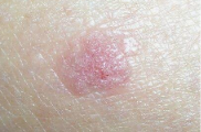
Early squamous cell carcinoma (SCC) usually looks like a persistent scaly red patch, a firm red nodule, or a crusted sore that won't heal after 3–4 weeks. It grows over weeks to months on sun-exposed skin — face, ears, scalp, lips, backs of hands — and can bleed or ulcerate. Any non-healing rough patch should be reviewed by a dermatologist within 2–4 weeks. SCC caught early is almost always curable.
TL;DR: Key Takeaways
- Persistent scaly red patch, firm nodule, or non-healing sore on sun-exposed skin
- Grows faster than BCC — weeks to months, not years
- 2–5% metastasise if untreated; risk is higher on lip, ear, or in immunosuppressed patients
- Cumulative UV exposure is the main cause — tanning beds raise risk ~70%
- Any rough spot that thickens, ulcerates or bleeds → dermatologist within 2–4 weeks
At a glance
Typical appearance
Rough, scaly, red or crusted; sometimes wart-like or ulcerated
Typical age
50+, fair-skinned adults with cumulative sun damage
Growth speed
Weeks to months (faster than BCC)
When to check
Any sore that doesn't heal in 3–4 weeks — dermatologist within 2–4 weeks
What is Squamous Cell Carcinoma?
Squamous cell carcinoma (SCC) develops in the squamous cells that make up the middle and outer layers of your skin. It's the second most common skin cancer after basal cell carcinoma, with over 1 million cases diagnosed annually in the US.
Unlike BCC, SCC can be more aggressive and has the potential to spread to lymph nodes and other organs if left untreated. This is why early detection matters — a small SCC caught early is almost always curable with simple surgery.
Warning Signs of SCC
See a doctor within 2–4 weeks if you notice:
SCC vs BCC: Key Differences
SCC
- • Grows faster (weeks–months)
- • Can spread to lymph nodes
- • Often rough, scaly texture
- • Higher risk in immunocompromised
BCC
- • Grows slowly (months–years)
- • Rarely spreads
- • Often pearly, waxy appearance
- • Very low metastasis risk
For a deeper visual comparison with the precancerous stage, see SCC vs Actinic Keratosis.
Risk Factors
What to do if a spot looks like SCC
- Photograph the spot with a ruler or coin for scale — document the date
- Run a free AI screening as a first triage step
- Book a dermatologist within 2–4 weeks if the spot has not healed
- Do not pick, scrape, or burn suspicious lesions — this can delay diagnosis
- Bring your photos to the appointment so the clinician can assess change over time
Frequently Asked Questions
Early SCC often appears as a persistent, scaly red patch, a firm red nodule, a flat sore with a scaly crust, a new growth or raised area on an old scar, or a rough, thickened patch on the lip. It may resemble a wart or non-healing sore.
SCC is more aggressive than basal cell carcinoma. While most are curable with early treatment, SCC can spread to lymph nodes and distant organs if left untreated, especially in high-risk locations like lips, ears, or in immunocompromised patients.
SCC typically grows faster than BCC—over weeks to months rather than years. Rapid growth is more common in SCC, and this faster progression is one reason early detection matters even more.
The main cause is cumulative UV damage from sun or tanning beds. Other factors include fair skin, older age, weakened immune system, chronic skin inflammation, HPV infection, arsenic exposure, and previous radiation treatment.
SCC commonly occurs on sun-exposed areas: face, ears, scalp, neck, lips, back of hands, and forearms. However, it can also develop on areas not exposed to sun, including genitals, inside the mouth, and in chronic scars or wounds.
Actinic keratosis (AK) is a precancerous condition—rough, scaly patches from sun damage that can progress to SCC. About 10% of AKs become SCC. SCC is actual cancer, while AK is a warning sign requiring monitoring or treatment.
Unlike melanoma, SCC does not typically develop from moles. It arises from squamous cells in sun-damaged skin. SCC often develops in areas of chronic sun damage, actinic keratoses, or in old scars and chronic wounds.
Treatment options include surgical excision, Mohs surgery (especially for facial SCC), curettage and electrodesiccation, cryotherapy for small superficial lesions, radiation therapy, and topical/systemic medications for certain cases.
High-risk SCCs include those larger than 2cm, poorly differentiated (aggressive cell type), deep invasion, location on lips/ears/temples, arising in scars or chronic wounds, and occurring in immunocompromised patients. These require more aggressive treatment.
Yes, unlike BCC, SCC can metastasize. About 2-5% of SCCs spread to lymph nodes or distant organs. Risk of spread is higher with aggressive subtypes, large tumors, deep invasion, and certain locations like lips and ears.
Lip SCC often appears as a persistent rough, scaly patch that doesn't heal, a thickened area, a sore that crusts or bleeds, or a warty growth. It commonly occurs on the lower lip due to sun exposure and may be mistaken for chapped lips.
Prevention includes: daily SPF 30+ sunscreen, protective clothing and wide-brimmed hats, avoiding peak sun hours, never using tanning beds, protecting lips with SPF lip balm, and having regular skin checks if high-risk.